top of page
Search

Jacobs Endodontics | One Year On

  • Rob Jacobs
  • Apr 23
  • 3 min read

Thank you very much to all dental professionals that have referred their valued patients to Jacobs Endodontics - one year has flown by!


It has been a really rewarding and enjoyable experience continuing to enjoy clinical Endodontic work, whilst also nurturing and building the business.


Below are 2 interesting cases where I have carried out a detailed review and reflection, whcih I hope is of interest to you.


Case 1: Upper Left First Molar – Complex Canal Anatomy


A 38 year old female patient was referred for endodontic treatment of her upper left second premolar and upper left first molar.


The general dentist had removed caries in both teeth and placed biodentine interim restorations trying to maintain vitality of the teeth. Unfortunately, the two teeth became symptomatic.


A CBCT scan revealed complex anatomy in both the DB and MB roots of the UL6.


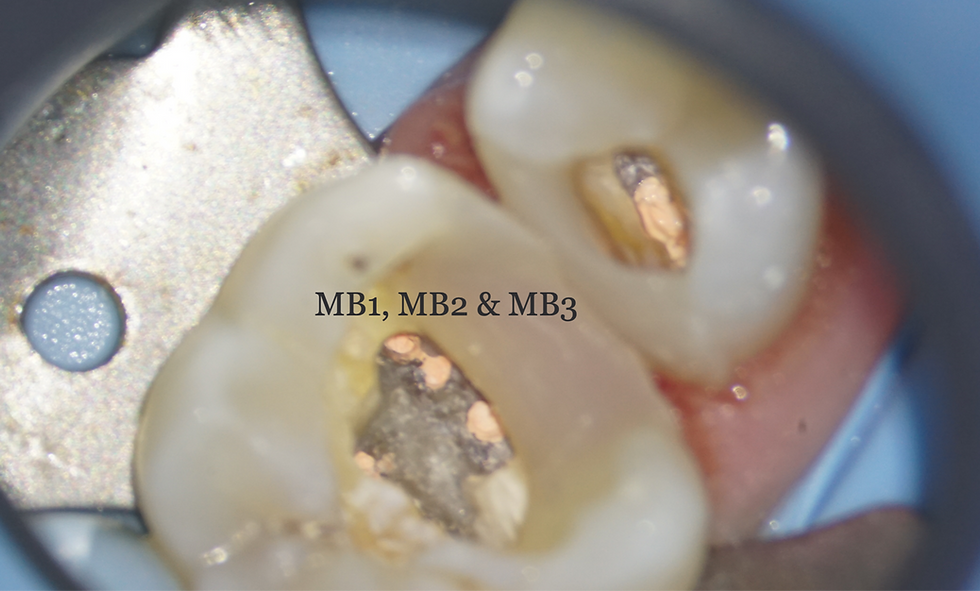
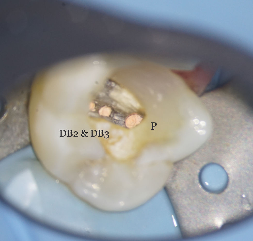
During treatment of the UL6, careful exploration under magnification revealed a highly complex canal system, with seven canals identified and managed!
Once again the axial view of the UL6 showcases the anatomy of the root canal system.
Presenting situation biodentine breaking down as placed a few months earlier
All biodentine removed, pulps accessed and covered with PTFE, walls rebuilt with sectional matrix bands and SDR Flow + Composite from Dentsply.
3 DB canals following preparation

Sealer placement showing confluence of the DB canals. The attached videos showcase a snippet of the Radio 2 quiz which is often our morning audio choice in surgery!


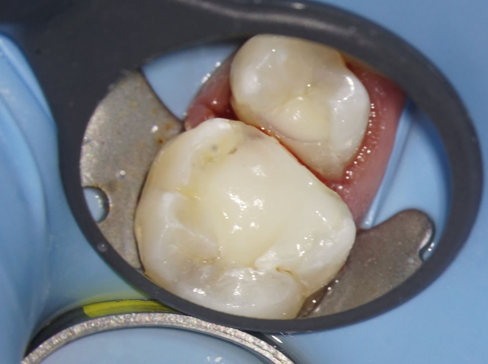
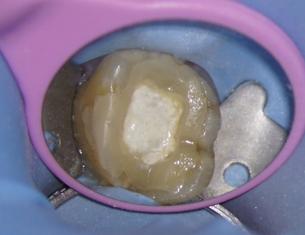
Dual-cure Composite cores (Luxacore Z Dual from DMG).

Conclusion: Additional canal anatomy in maxillary molars is not uncommon. A systematic approach—supported by magnification and appropriate imaging—remains key to identifying and managing these cases predictably.


Case 2: Upper Left First Molar – Fundamentals for Success


A 29 year old male patient was referred for endodontic treatment of his upper left first molar.


The general dentist had extirapted the tooth but was having difficulty completing the case and had separated an instrument coronally in the MB root.


In this case, the key factor was to dismantle the previous restoration and fully unroof the pulp chamber, allowing removal of the fractured instrument, location of the 4 canals, and predictable shaping, disinfection and obturation of the root canal system.
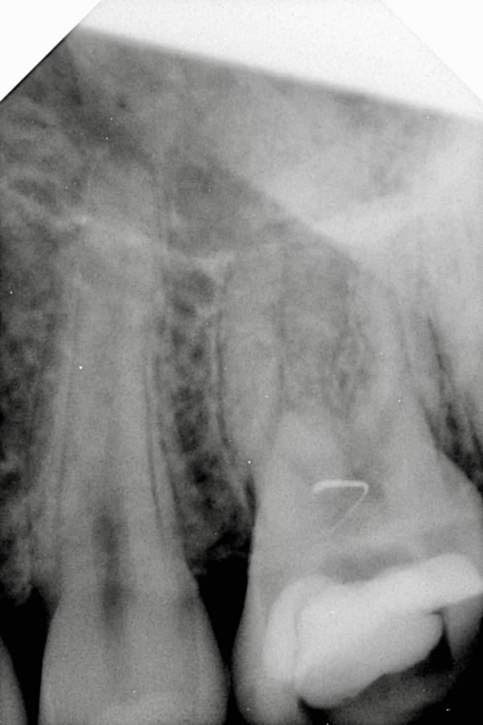
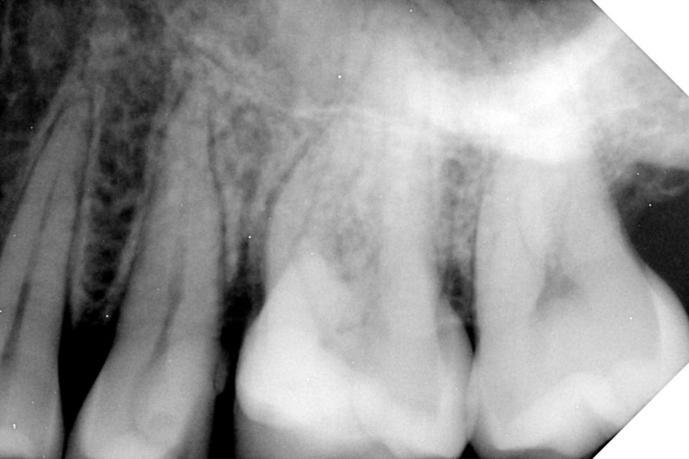
Presenting situation. Multiple restorations and separated instrument in canal orifice
The axial view of the CBCT (bottom left image) shows the anatomy of the UL5 and UL6 in detail.
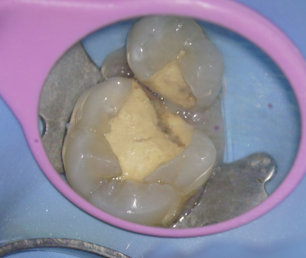
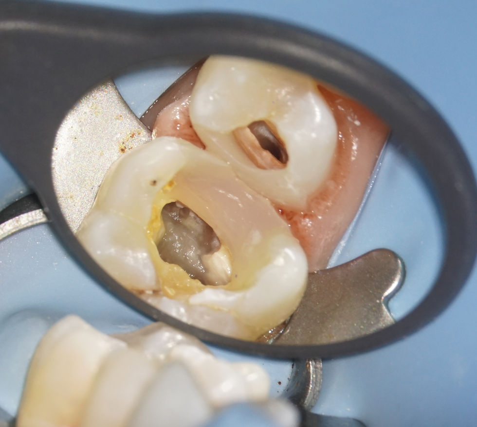
Multiple restorations - let's remove all the restorative and see what lies beneath!
Initial extirpation has only partially unroofed the pulp chamber, hence the difficulty in treating the tooth and the resultant file separation.
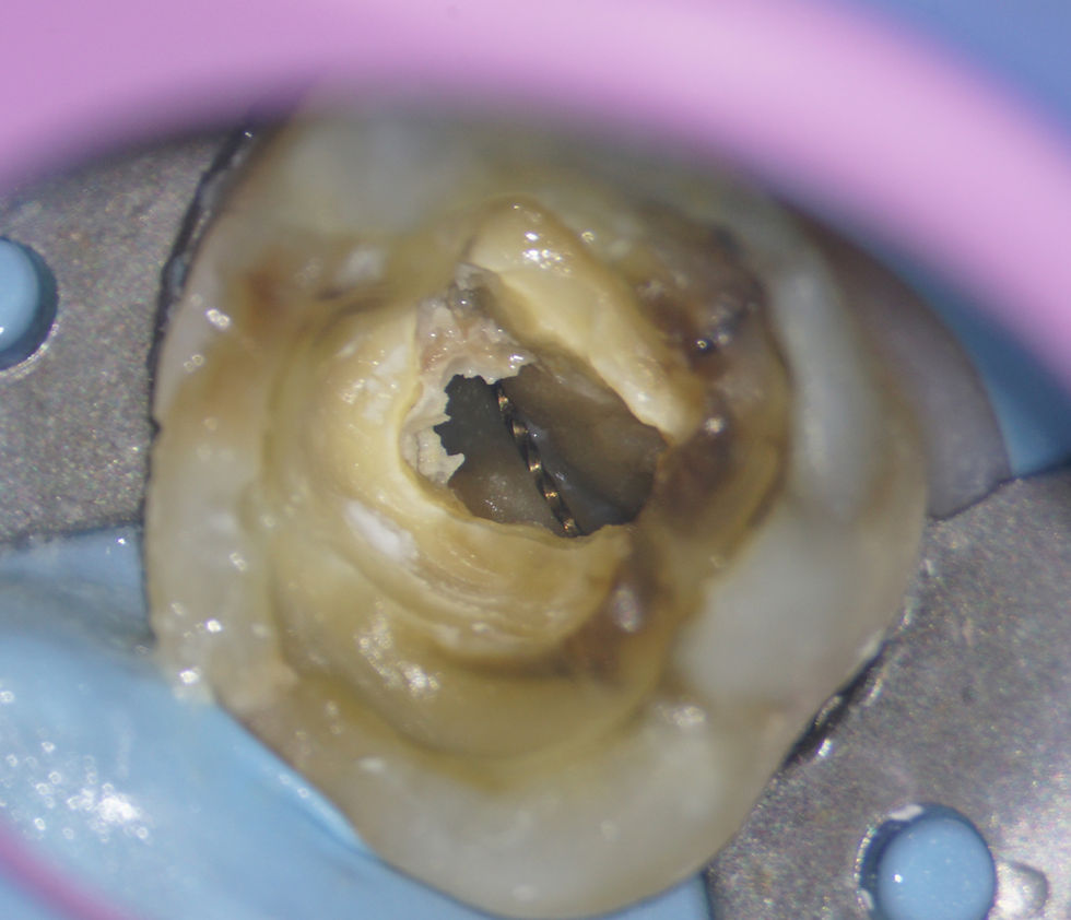
As the pulp chamber is unroofed, the tip of the Wave One Gold file begins to be uncovered and removal is straightorward with the use of ultrasonics (Satellec 10z tip).
MB2 located and prepared. Note the darker colour of the dentine on the pulp floor compared with the walls
The benefits of full restoration removal is that cracks can be visualised and a comprehensive assessment made of tooth longevity / restorability.
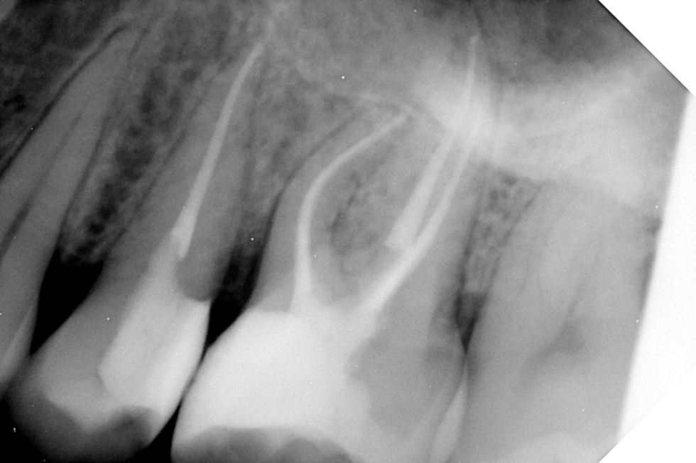
Obturated case ready for core build up
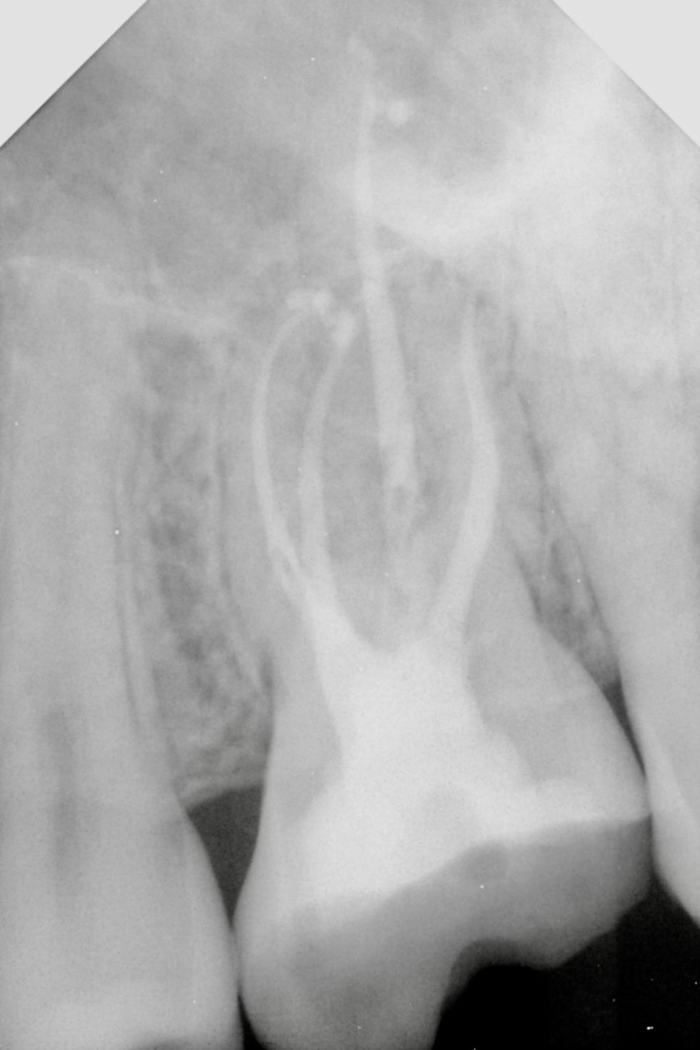
Four canals filled to length, well sealed at all apices with a small sealer puff.

Conclusion: Unroofing the pulp chamber fully is key to allowing full inspection and location of the root canal anatomy. This key step allows the file removal, preparation and obturation to be striaghtforward.


Comments


©2025 by Jacobs Endodontics.

bottom of page